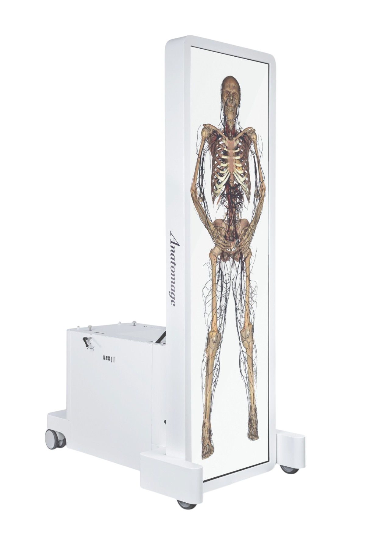
Flexibile and effective educational tool
The Table provides a learning-by-doing experience to everyone, erasing the difficulty of traditional teaching methods many students face.
Impacted research proves the interactive experience provided by the Table is more engaging than the experiences provided by traditional learning tools.
The ability to use the Anatomage data from anywhere makes any online or on-site lesson more accessible.
Instructors can explain complex topics, save, and share contents in a few steps.
Students can revise the contents using the Table, alone or in a group, before or after the lesson.
ULTIMATE VASCULAR TRACING
The cardiovascular system can be explored with an unprecedented level of accuracy.
A new algorithm has been implemented to identify even the smallest vessels of the pre-existing bodies.
Users can toggle structures on and off, identify names and functions of the structures, and better comprehend intricate areas of the body through the kinematic visualisation of heart beating and blood flow in all the different tracts of the cardiovascular system.
INTERACTIVE DISSECTION
The Table offers unique interactive dissection and reference tools.
Users can rotate the body, cut in any plane and visualise the name of each displayed structure.
A dynamic view of internal anatomy is runtime reconstructed with vivid life-like colours to get a realistic feeling of each structure and the surrounding ones.
Different scalpels are available to perform linear cuts on the anatomical planes, or free-hand cuts to remove skin, subcutaneous fat, and deeper structures in a specific region.
Cuts can be used as an educational tool to mimic dissection, or to simulate a surgical procedure with the eventual insertion of a medical device.
The possibility to undo at any time, makes the bodies dissectible for an endless number of times.
IMPRESSIVE 3D RECONSTRUCTIONS
Anatomy learning reaches an unparalleled level of understanding when students can compare digital datasets of prosections with frozen bodies datasets.
The Table contains 66 high-quality surface scans of prosections.
The latest additions provide users with 3D data showing different dissection stages.
The surface has been mapped during each stage to highlight and identify structures.


