


The full anatomy of four animals (a dog, a cat, a frog and a mouse) is volumetrically displayed from head to toe and it is directly connected to the picture of each slice. The vivid colours and shape of the bodies are preserved to accurately depict real anatomy. The digital animal bodies can be explored with dissection cuts layer by layer, re-vealing the details of internal structures.

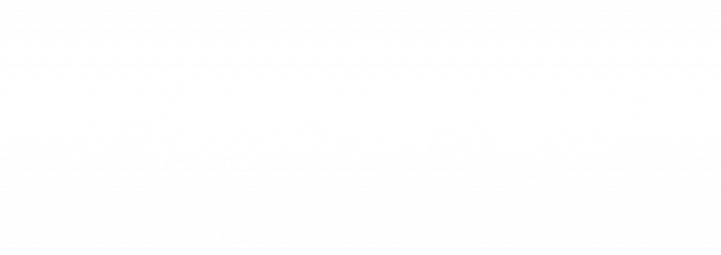
As the world’s first exceptionally accurate dog anatomy atlas, Anatomage Dog features comprehensive annotated internal organs, including vascular systems and muscoloskeletal structures, as well as more than 1100 anatomical structures segmented, and 3D layer-by-layer dissection capabilities.
The Table Vet comes with a diverse anatomy case library with 285 annotated animal cases and pathology reports. Users can learn how to identify abnormal anatomy – including suspicious masses – that stem from potential carcinoma or fungal infection.
HIGH ACCURACY AND TRUE TO LIFE VISUALS
A plethora of DICOM cases from various animal species with unrivaled accuracy, high-quality renderings are a welcomed addition to Table Vet.
The ability to reconstruct DICOM data in real-time and apply the numerous different renderings to each scan provides the user with limitless learning opportunities.
Users can go one step further by loading individually and previously acquired DICOM scans to the Table Vet, all the while enjoying the same high-quality interactive teaching experience.
Users can expedite their diagnosis process by comparing their data with the pre-existing data available within the Table Vet.
PHOTOREALISTIC ANIMAL ANATOMY
As Anatomage Table Vet contains real-tissue animal bodies, it offers an unprecedented learning experience for those who would like to interact with real animal cadavers in a chemical-free, technologically inspired space.
With Anatomage’s renowned 3D visualization capabilities, Anatomage Table Vet allows students to visualise and study the animals’ internal anatomy, one step forward from the limited availability of deceased animal bodies.
VIRTUAL DISSECTIONS
Over 10 years in the making, Anatomage Tables offer unique interactive dissection and reference tools.
Users can rotate the animal bodies and cut on any plane.
A dynamic view of internal anatomy is runtime reconstructed with vivid life-like colours to get a realistic feeling of each structure and the surrounding ones.
Different scalpels are available to perform planar cuts on the anatomical planes, or free-hand cuts to remove skin, subcutaneous fat, and deeper structures in a specific region.
The possibility to undo at any time makes the bodies dissectible for an endless number of times.
ANNOTATIONS, QUIZZES & CURRICULUM PREPARATION
For the first time, Table Vet offers the possibility to visualise the full segmentation of the dog and its annotated structures.
All anatomical structures can also be highlighted with pins to create customised quizzes.
Interactive automated quiz modes are also available to engage the students more than ever.
Thousands of presets can be saved, exported and recalled anytime.
Each preset helps the user in identifying the main points from each lesson.
Each visualisation can be exported as an image or a video.